Is fluorography harmful and why is this x-ray examination method prescribed? The result is an image of the internal organs.
It can be projected onto a special film or converted into digital format. Due to radiation, the method is not absolutely safe, but to assess how harmful fluorography is, you need to understand all the features of the examination.
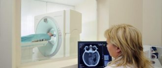
Read how MRI of the brain is harmful to your health -
Concept and types
Fluorography is a special technique that helps determine the condition of the chest organs.
The procedure is carried out using X-ray waves and is based on the different abilities of organs to transmit radiation. The test result appears on the screen of the specialist, and a study is carried out on it. Fluorography plays the role of prevention. It is not possible to make an accurate diagnosis and prescribe treatment using an image; it is used as an additional examination. However, an experienced physician can easily determine the presence of pathological changes and refer you to a specialized doctor.
There are two types of chest examination. The harm of the procedure is determined by the chosen type of inspection.
This type has been used for a long time. X-ray beams are passed through the patient's chest from the back. A special film produces an image of organs and bones. To obtain an image, you need to develop the film in a special solution. This is one of the disadvantages of the procedure.
This method is considered harmful; the person receives a higher dose of radiation than with the procedure using new technologies. With film fluorography, the patient receives a dosage equal to half the permissible value per year.
Research defects occur in fifteen percent of all cases. Repeating the method leads to an increase in the dose of radiation received. Currently, many medical institutions are trying to abandon this harmful technique.
This is a new method of fluorography. Special rays are passed only through organs that require examination. The finished image appears on the screen, then it is examined by a specialist. The device allows you to get results quickly. The procedure has advantages that make it popular in many hospitals.
Pros:
- High accuracy of results,
- The original data is present on the computer, so no repeated checks are required,
- No film or developing materials required
- The received information can be stored on removable media,
- Doesn't require a lot of money
- The method allows you to check many people.
Digital fluorography is popular and not harmful compared to the film method.
Survey results
If the tissue density in the examined organs is changed, this will be noticeable in the resulting image. Often, fluorography reveals the appearance of connective fibers in the lungs. They can be located in different areas of the organs and have different appearances. Depending on this, fibers are classified into scars, cords, fibrosis, adhesions, sclerosis, and radiance. Cancerous tumors, abscesses, calcifications, cysts, emphysematous phenomena, infiltrates are also clearly visible on the images. However, the disease cannot always be detected using this diagnostic method. For example, pneumonia will be noticeable only when it takes on a fairly advanced form.
The fluorography image does not appear instantly, it takes some time, so the results of the examination can only be obtained within a day. If no pathologies are found, the patient is given a stamped certificate indicating this. Otherwise, a number of additional diagnostic measures are prescribed.
What is the harm from fluorography to humans?
Is chest x-ray procedure harmful to health? The greatest harm from fluorography is radiation. There is a concept of effective equivalent dose. The number indicates possible risks and the development of complications after the procedure.
For film fluorography, the indicator varies from 0.5 to 0.8 mSv. If the examination is carried out using digital equipment, then the EED is 0.04 mSv.
The difference is big, but not all government institutions have the opportunity to choose the type of research. Often budget organizations use old equipment.
It is recommended to understand that x-rays and fluorography are different techniques. With X-rays, the image is clearer and the results are more accurate. But the radiation and harm in such a case are higher. Therefore, it is not recommended to go for an x-ray without a doctor’s prescription.
In what cases is examination of the chest organs using fluorography prescribed? There are groups of people who are recommended to undergo the procedure at least once a year.
Groups of people:
- Patients with the possible development of diseases of the respiratory, genitourinary systems, pathological processes in the endocrine system, with ulcerative lesions of the stomach and duodenum.
- In people undergoing radiation, cytostatic and steroid treatment.
- Patients with no fixed place of residence,
- People working with babies and adolescents.
- Employees of medical institutions, sanatoriums, sports and secondary schools.
Some people require screening twice a year. Such research cannot be abandoned to avoid negative consequences.
Category:
- Presence of HIV infection,
- Previous tuberculosis (first three years),
- Release from prison (first two years),
- After close contact with carriers of the Koch bacillus,
- Workers at tuberculosis clinics and maternity wards.
They must undergo additional examination if they suspect the presence of tuberculosis, unpleasant symptoms in the respiratory tract, HIV infection, during military conscription, or if there is a pregnant woman in the house.
It is possible to refuse fluorography, but it is recommended to think about the consequences.
How often are diagnostics allowed?
This question worries many. For preventive purposes, tuberculosis should be examined at least once every two years. People who have special indications should resort to this diagnostic method more often. For example, for those who have been diagnosed with tuberculosis in their family or work team, fluorography is prescribed every six months. Workers of maternity hospitals, tuberculosis hospitals, dispensaries, and sanatoriums are examined at the same frequency. Also, every six months, diagnostics are carried out for people with severe chronic pathologies, such as diabetes, bronchial asthma, stomach ulcers, HIV, and so on, as well as those who have served time in prison. For conscripts into the army and persons diagnosed with tuberculosis, fluorography is done regardless of how much time has passed since the previous examination.
What is the harm from fluorography to humans?
Is it harmful to have fluorography? The procedure performed harms the body with its radiation. However, if the examination is carried out using modern equipment, the negative impact is minimal. The harm from radiation increases with simultaneous research using other radioactive techniques.
There are positive aspects to such a survey. A timely procedure helps determine the development of inflammatory processes at the initial stage. For some diseases, fluorography is used to monitor the course of the disease.
Any person has the right to refuse the harmful effects of radioactive rays on his body. However, it is recommended to remember that the result in the future is unpredictable, and the risk of missing time to treat the disease increases.
Contraindications
Fluorography has no contraindications. Features are identified in which the examination is harmful and is not recommended.
Peculiarities:
- The inability of a person to remain upright,
- Fear of enclosed spaces, lack of air,
- Women during pregnancy, especially in the first trimester,
- While breastfeeding your baby,
- In children under fifteen years of age.
In many ways, the possibility of performing fluorography depends on the person’s condition.
Digital technology
Nowadays film technology is still used everywhere, but an advanced method has already been developed and is being used in some places, which has a number of advantages. Digital fluorography allows you to obtain the most accurate images, and at the same time the patient is exposed to less radiation. The advantages also include the ability to transmit and store information on digital media, the absence of expensive materials, and the ability of devices to “serve” a larger number of patients per unit of time.
Digital fluorography is more effective than film fluorography (according to some data) by about 15%, while at the same time, during the procedure, the radiological load increases five times less than when using the film version. Due to this, even children can be diagnosed using digital fluorograms. Today, there are already devices equipped with a silicon linear detector that produce an amount of radiation comparable to what we receive in one day during normal life.
Methodology
The patient is invited to the room for fluorography. The procedure is carried out in a standing position. For preventive purposes, images of the chest are taken in anterior projection. To do this, the patient is pressed with his chest against a fluorescent screen, inside of which there is a film cassette. The chin is placed in a special depression, the elbows are pulled back.
The patient takes a deep breath and holds his breath. At this time, an X-ray tube located behind it emits beams of radiation. Passing through the chest, some of them are absorbed, the other ends up on the photosensitive matrix. The image from the matrix is transferred to film. The whole process takes a few seconds.
To diagnose pathologies, photographs of the lungs taken from different angles may be required. In this case, the patient changes his body position several times, pressing against the screen first with his chest, then with his back and side.
Indications for the procedure
Fluorography is considered a screening test. This means that all adults in the country undergo it regularly in order to recognize the signs of dangerous lung diseases in time. Most employers require employees to undergo timely examinations, and those who do not do this are not allowed to work.
Fluorography allows you to detect in the early stages:
- tuberculosis;
- pneumonia;
- tumors;
- cysts;
- inflammatory changes;
- accumulations of gas and liquid;
- foreign objects.
For greater accuracy, each image is examined by two doctors - this helps to avoid mistakes. If necessary, more specialists can be involved.
Also, patients who have been admitted to a medical institution, intend to get a job, study or serve in the army, if their last fluorography was done more than a year ago, undergo mandatory analysis.
City Medical Center issuing certificates
Despite the variety of more modern methods for examining the lungs and other internal organs, fluorography is still the most popular. And thanks to modern fluorographic technology and the qualifications of specialists, periodic medical examinations make it possible to identify pathology quickly and accurately.
The great advantage of this type of radiation diagnostics is its low cost compared to such expensive services as ultrasound and MRI.
The information content of X-rays and fluorography for examining the chest is also higher than that of the above methods.
Plus, examination with a fluorograph is a mandatory procedure in all clinics. The goal is to identify possible hidden pathological processes.
If you urgently need to check your lungs, we recommend that you do fluorography for a fee at an affordable price at the Medical Driving Commission in Moscow . Here we organize examinations for FLG at the highest level, using the latest equipment and medical methods.
The essence of the procedure
What is this medical procedure? Fluorography is the photographic recording of patient tissue on a special film.
The image that appears on a special fluorescent screen during radiation is transferred to this film in miniature form.
Thanks to advanced digital equipment, images obtained through fluorography are sharp, clear, and can easily be used through a magnifying glass to make a diagnosis.
The advantage of conducting periodic medical examinations using a fluorograph is the relatively small dose of radiation received by the patient. Therefore, examinations can be carried out once a year, and, if necessary, twice a year: for those who have recently been discharged from correctional institutions or have suffered from serious illnesses.
Contraindications for fluorography
Although the patient receives minimal radiation during the procedure, radiation is still present.
The principle of the examination is also important - the patient, in a standing position, presses his chest against the screen to take a picture of the organs. Otherwise, the procedure cannot be carried out.
Therefore, there are several categories of people for whom fluorography is strictly contraindicated:
- pregnant and breastfeeding women;
- children under 16 years of age;
- bedridden patients or disabled people who are unable to stand;
- suffering from severe shortness of breath.
In what cases is it necessary to undergo a fluorographic examination?
It is necessary to undergo fluorography for a fee on an unscheduled basis if:
- You suffer from tuberculosis of any form;
- you suspect neoplasms (tumors, cysts) in the chest organs;
- you have an inflammatory process in the lungs (pneumonia, pleurisy, bronchitis);
- You suffer from cardiovascular diseases.
For everyone else, undergoing fluorography in Moscow and other cities of the Russian Federation is mandatory at least once a year as a preventative measure. These categories of patients include:
- all people over 16 years of age;
- those living with pregnant women and infants;
- young people drafted by the military registration and enlistment office;
- HIV-infected.
A routine check of organs on the device is necessary for the timely detection of hidden inflammatory and cancer processes occurring in the body. Often, the patient may not even be aware of the disease, since the symptoms do not appear outwardly. Only with the help of a professional fluorograph can negative changes in the body be identified and treatment prescribed in a timely manner.
Source: https://www.medspravka.org/articles/protivopokazaniya-dlya-provedeniya-flyuorografii/
Story
Wilhelm Conrad Roentgen discovered a special type of radiation in 1895, completely by accident, as is usually the case with great discoveries. Finishing his experiments, one November evening, he noticed the appearance of a strange glow of a substance left on the table. The substance in the glass jar glowed due to the turned on electron vacuum tube, which Roentgen simply forgot to turn off.
The year 1901 was marked by the awarding of the Nobel Prize in physics for this truly significant, great discovery. This is how a separate field appeared in medicine – radiology.
Today, doctors all over the world have such a wide range of X-ray examinations that in the days of Roentgen it was not even possible to dream of this: various methods of X-ray, fluoroscopic examinations, native and using contrast agents, computed tomography examination, screening research methods (for example, fluorographic examination ). There is no area of modern medicine today where X-ray diagnostic methods are not used. Today, the simplest and most common method of x-ray examination, which we all encounter regularly, is fluorography (FLG).
Exposure to ionizing radiation
Ionizing radiation has found wide application in our everyday life: industrial production, agriculture, scientific research, and medicine. Each of us in the process of life is exposed to natural radiation (water, soil), or artificial (for example, from medical sources).
The discovery of X-rays is, of course, important for industry and medicine, but it also poses a potential danger if used incorrectly, exceeding the maximum permissible doses, or unreasonably used.