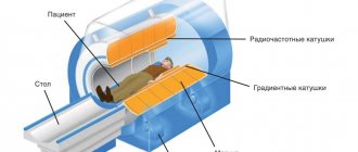
The principle of operation of the CT device is circular layer-by-layer scanning of the examined area with a thin beam of X-rays. The structures of the human body have different densities, so they absorb X-rays with varying degrees of intensity. The beam attenuation parameters are recorded by a system of detectors installed on the opposite side of the emitter and converted into electrical signals.
The dose of radiation received by a patient during a CT scan of the brain is 100-500 times higher than that typical for classical radiography. The exact value depends on the model of the device, the installed operating mode of the equipment and the diagnostic complexity of the study. The effective radiation exposure for a head CT scan is approximately 2-4 mSv, which is similar to the exposure a person receives from background radiation over 2-5 years.
The main limitation to the brain CT procedure is pregnancy, since there is a potential risk of developing defects in the fetus when exposed to X-rays. However, pregnancy is not an absolute contraindication - CT can be performed in exceptional cases, when the benefit of diagnosis outweighs the possible harm or in emergency cases. It is also not recommended to perform a CT scan of the head in children under 14 years of age unless there are serious clinical indications.
Let us note that for 98-99% of patients who performed this examination, there are no negative consequences of brain CT, the procedure does not harm health and turns out to be absolutely safe.
How CT works
Examinations using computed tomography scanners have been carried out for about forty years, characterized by speed, moderate cost compared to MRI, and wide diagnostic capabilities.
They are used to study functional and anatomical changes in:
- heart and blood vessels;
- brain;
- musculoskeletal system;
- lungs and gastrointestinal tract;
- liver and kidneys;
- organs of the reproductive system;
- bladder;
- thyroid gland, etc.
CT is based on the use of X-rays, which pass through organs, bones, muscles, cartilage and other tissue at different speeds, forming a clear image.
The main structural elements of the tomograph are a movable table and a ring-shaped installation (gantry) on which sensors are fixed (the number depends on the type of device). They receive the return signal, transmit it to the analyzing unit, which decrypts the data, after which an image is created in increments of 1-5 mm.
Depending on the suspected or already identified pathology, you will be prescribed a certain type of CT scan: multislice tomography, enhanced procedure, DSCT, CT angiography or CT perfusion.
The cost of diagnostics will vary depending on the clinic where you decide to go, the parameters of the device, the examination area, and the use of a contrast agent.
Contrast agent for tomography
Contrast agents are needed to “highlight” certain structures on x-rays.
The contrast agent blocks X-rays and lights up white in pictures, which helps highlight blood vessels, intestines or other parts of the body. This will enable the doctor to recognize certain defects that would go unnoticed without contrast. There are several ways to administer a contrast agent:
• Oral. If the doctor plans to examine the esophagus or stomach, then the contrast must be taken in the form of a special solution. It may have an unpleasant taste. • Parenteral. Contrast agents are often injected into the bloodstream through a vein in the arm. This is done before studying the gallbladder, urinary tract and blood vessels. During the injection, the patient may experience a feeling of heat at the injection site, as well as a metallic taste in the mouth. • Rectally. To visualize the intestine, a contrast agent is administered rectally. This procedure is associated with discomfort. There must be enough contrast agent so that it covers the intestinal walls and makes them clearly visible on the pictures.
Preparing young children for CT scans is very challenging. Before the scan, your doctor may recommend a sedative to help your child remain quiet and not twitch during the procedure. Any, even minor movements can affect the results of a CT scan because the clarity of the images is lost.
Are there any harms from having a CT scan?
Having received an appointment for a computed tomography scan, patients often wonder whether it is worth doing it, given the potential harm to the body.
However, provided that the permissible radiation dose is observed, there is no likelihood that CT will cause the development of oncology or other diseases.
Modern models of computed tomographs have significantly less radiation exposure than those that were used before.
Therefore, if you are examined in a medical center with the latest generation equipment and only when necessary, then the risk of critical consequences is minimal.
The degree of exposure is determined by two main factors:
- characteristics of the device;
- size and specificity of the scanning area.
To study the condition of the abdominal organs, pelvis, lungs and chest, the most powerful X-ray irradiation is required, but when examining the brain or limbs, the radiation dose is one and a half to two times less.
None of the clinical studies conducted by scientists have proven that tomography provokes the development of oncology and other disorders.
Attention! Cancer and other negative consequences can occur only if there are initial prerequisites, for example, weakened immunity, chronic inflammatory processes, untreated infections.
The advisability of a CT scan is determined by the doctor who is treating you, based on your general health, diagnosis, etc.
How is CT more harmful than MRI?
In order to answer the question of what is more harmful than MRI or computed tomography, it is necessary to consider the principle of the design and operation of the devices of such examination methods.
When examining a patient with a computed tomograph, his body is exposed to small doses of ionizing radiation. When this procedure is repeated, the radiation accumulates, so it has some limitations and contraindications . For example, examination in this way is prohibited for pregnant women.
In diagnostic devices based on MRI, there are no sources of radiation, and the patient during the examination is exposed only to a strong, but completely safe electromagnetic field. Therefore, with such devices it is possible to undergo repeated examinations without restrictions.
How many times a year can a CT scan be done?
Due to the fact that CT involves contact with X-rays, it should not be performed more often than necessary. The number of times per year is determined by the radiation dose, but the optimal option from a health point of view is 1 procedure every 12 months.
If we are talking about emergency medical indications, then scanning is performed up to 4-5 times a year, and the interval between examinations should be at least a month.
The established norm of 150 mSv per year is relevant for employees of x-ray rooms. It is believed that such an amount of radiation is not capable of producing obvious negative consequences for the functioning of internal organs and systems.
Side effects of positron emission tomography
The state of health after PET/CT is not disturbed or disturbed, however, with some diseases it is difficult for the patient to remain motionless for a long time. The most common side effects of the diagnosis are weakness and dizziness due to dietary restrictions and the duration of the study. The administered medications are non-toxic, prepared under sterile conditions and meet the requirements of state standards. If you follow your doctor's recommendations, PET/CT has no negative consequences.
A 19-year-old female patient was diagnosed with diffuse large B-cell lymphoma. The image on the left is the results of a PET/CT study to assess the extent of the malignant process (red areas) before treatment, on the right are the results after two courses of chemotherapy.
Contraindications for CT scanning
A CT scan has many advantages (efficiency, information content, possibility of use in emergency situations), but it is not safe for all patients. The procedure is not performed if:
- pregnancy and lactation period in women;
- the patient’s weight is over 140-150 kilograms (explained by the physical capabilities of the device);
- renal dysfunction and allergic reaction to contrast during enhanced examination;
- claustrophobia and mental disorders;
- diseases and pathological conditions that do not allow keeping the body in a stationary position;
- the presence of plaster or metal structures in the area under study;
- multiple myeloma.
Important! In particularly severe cases, a specialized specialist may prescribe a CT scan even in the presence of relative contraindications, if its benefits outweigh the harm to health.
A referral for tomography is not formally required, but the body should not be exposed to radiation unless the attending physician insists on it.
In addition, it is better to come to the scan with the results of other tests and studies, as well as a preliminary diagnosis made by a gastroenterologist, neurologist, oncologist, orthopedist, etc.
CT of the brain CT of the abdominal cavity CT of the sinuses CT of the kidneys CT of the lungs CT of the pelvis CT of the knee joint CT of the hip joint CT of the heart CT of the cervical spine CT of the thoracic aorta CT of the shoulder joint CT of the elbow joint CT of the thoracic spine CT of the lumbosacral spine CT of the temporal bones CT of the facial bones CT of the adrenal glands CT of the abdominal aorta CT of the brain vessels CT of the jaw CT of the orbits CT of the teeth CT of the neck vessels CT of the chest CT of the foot CT of the pelvic bones CT of the hand CT of the vessels of the lower extremities CT of the intestine CT of the larynx CT of the bladder CT of the ankle joint CT of the coccyx CT of the coronary vessels CT of the soft tissues of the neck CT of the skull CT of the thyroid gland CT of the liver CT of the wrist joint CT of the inner ear
Informativeness and safety of positron emission tomography
PET/CT is the optimal examination method, as it combines positron emission tomography, which helps to see changes at the cellular level, and computed tomography to assess the structure of the organ. As a result of the diagnosis, the doctor receives more complete information about the pathological process; tumors from 6 mm in greatest dimension are visible in the images.
Positron emission tomography allows you to determine the stage of the disease and plan treatment, reduce the radiation dose to surrounding healthy tissue during radiotherapy by accurately determining the location of the tumor. It is indispensable in assessing the prevalence of certain malignant neoplasms, such as lymphoma, melanoma and others.
see also
- What is echogenicity?
- Biochemistry analysis: how to prepare
- Fetal weight by week
- How long does it take for an ultrasound to show the gender of a baby?
- What not to eat before testing for occult blood
- Few red blood cells in the blood, what does this mean?
- Normal level of alkaline phosphatase in the blood in men
- Normal prostate volume
- Blood test for TSH T3 and T4 readings interpretation
- The size of the prostate gland in men is normal with ultrasound
- Elevated ESR blood test
Indications and contraindications
CT scans are prescribed for various conditions to identify a pathological process or clarify the diagnosis:
- diagnosed cancer, metastases or suspicion of a malignant process;
- frequent, prolonged headaches without an obvious cause;
- problems with cerebral circulation and accompanying consequences;
- attacks of seizures, convulsions, loss of consciousness;
- conditions associated with experienced trauma;
- inflammatory processes localized in various parts of the body.
The benefits of computed tomography are undeniable - it helps to examine in detail almost any organ. In addition, this diagnostic method allows you to clarify the pathology identified as a result of other studies.
Among the contraindications, in which case it becomes dangerous to conduct this study, the following can be identified:
- syndrome of impairment of all renal functions;
- patients weighing more than 150 kg;
- applied plaster or metal structure in the examined area;
- claustrophobia (fear of closed spaces);
- pregnancy (especially in the first trimester);
- violent behavior of the patient against the background of mental disorders.
How often can the study be carried out?
PET/CT is performed at the direction of the attending physician as many times as the doctor deems necessary in each specific case. If it so happens that the patient is often subjected to various research methods, such as CT, radiography or fluoroscopy, fluorography, osteoscintigraphy, SPECT, you need to warn the doctor in advance before prescribing PET/CT.
Three treatments per year are usually safe for a person. It is advisable to conduct them no more than once a quarter in order to minimize harm from PET/CT examinations. If there are vital indications and on the recommendation of the attending physician, the examination may be carried out more often.
See also: Frequently asked questions about PET/CT
Difference in radiation level between X-ray and CT
Radiography is an inexpensive, informative way to visualize hard tissue. The information content of the examination is lower than computed tomography. Differences in tissue exposure levels require radiography to be performed first. If diagnostic confidence is not achieved, a CT scan is performed.
There are many types of radiography techniques. Examinations without contrast result in low radiation exposure. The level increases significantly when using fluoroscopy. The procedure involves studying the passage of barium through the esophagus and intestines. To reduce harm, the patient is protected by heavy lead shields. Sometimes radiography and fluoroscopy are performed simultaneously to reduce the amount of time that internal organs are scanned.
Despite the high ionization numbers after fluoroscopy, the radiation dose during computed tomography is significantly higher.
Is it dangerous to have an MRI while pregnant?
A woman’s condition, such as pregnancy, is a direct contraindication to CT scanning. It is recommended to discuss other testing methods with your doctor, such as MRI or ultrasound.
This procedure is not recommended before 12 weeks of pregnancy. In the first 3 months, vital organs form in the fetus. It is at this time that the unborn baby is most susceptible to the negative influences of the environment. The only exception in which an MRI is performed before 12 weeks is if there is a suspicion of fetal pathology. In other cases, the study is postponed to the 2nd or 3rd trimester of pregnancy.


