External and internal exposure
When assessing the effect of radiation on the body, it is necessary to distinguish between external and internal exposure.
External exposure
- exposure to ionizing radiation on the body from sources outside the body. In radiation safety engineering, it is accepted that external irradiation is created by sealed radioactive sources, for which the possibility of radioactive substances entering the environment under operating conditions is excluded. These include neutron sources, a-, b-, g-sources with a protective coating or shell, X-ray installations, and particle accelerators. Human exposure to penetrating radiation from nuclear explosions, cosmic radiation, and radiation from naturally occurring radioactive substances also constitutes cases of external exposure.
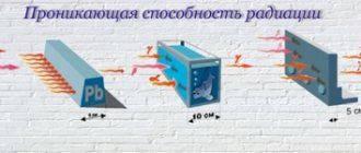
The degree of radiation damage to a person during external irradiation is largely determined by the penetrating ability of radiation, which depends on its type and energy. External irradiation with a-particles is not dangerous, since they penetrate into the outer layer of the skin (epidermis) only a few tens of microns and practically do not reach the sensitive layer of the skin (see table).
Beta particles penetrate several mm into the tissue, being absorbed by the skin and subcutaneous fat. When exposed areas of the body are irradiated with relatively large doses of b-radiation, skin erythema, eye cataracts, etc. may occur.
More dangerous is external γ-radiation, which permeates the entire body and can affect all human organs and tissues. This applies to an even greater extent to fast neutrons, which have high penetrating power and form recoil nucleus protons with high LET in tissue.
Internal exposure
– exposure of the body to ionizing radiation from radioactive substances located inside the body. Radioactive substances can enter the body through the respiratory tract, digestive tract, and through skin lesions. The danger of radionuclides entering the body arises when working with open sources and during radioactive contamination of the environment.
Let us consider the factors influencing the biological effect of internal irradiation.
• Activity
radionuclide in the body or critical organ. It is obvious that with increasing activity the degree of radiation damage increases.
• Type of radiation
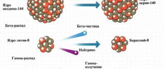
and its LPE. With the same activity of incorporated nuclides, the danger of internal exposure increases in the reverse order of external exposure: g-radiation < b-radiation < a-radiation (the most dangerous).
This is explained by the fact that particles are emitted and absorbed in the organs and tissues themselves, and their quality coefficient k plays the main role.
LET-dependent. Under the influence of a-particles, almost all cells near the a-source are destroyed, and a dense lesion is formed in the tissue. Cells affected by b-radiation are more dispersed in the tissue, which does not cause such a destructive effect; g-radiation ionizes through secondary electrons (indirect ionization) and, despite the same LET, creates much fewer ions in the tissue than b-radiation; In addition, part of the g-radiation energy goes outside the body. Therefore, other things being equal, internal irradiation with g-photons is the least dangerous.
• Localization
– the ability of some nuclides to selectively accumulate in individual organs of the body, called “critical”.
Critical organ -
an organ, tissue, part of the body or the entire body, the irradiation of which under given conditions causes the greatest harm to the health of the person or his offspring.
In descending order of radiosensitivity, three groups of critical organs are distinguished: Group I -
the whole body, gonads and red bone marrow;
Group II –
muscles, thyroid gland, adipose tissue, liver, kidneys, spleen, gastrointestinal tract, lungs, eye lens and other organs, with the exception of those belonging to groups I and III;
Group III –
skin, bone tissue, hands, forearms, ankles and feet. For example, about 20% of iodine is deposited in the thyroid gland, which by weight makes up only 0.03% of body weight. Sources of a-radiation - radium, uranium, plutonium and b-radiation - 90Sr, 90Y, 45Ca are deposited in bone tissue; 32P accumulates in the bones and lungs, 35S - in the lungs and gonads, 137C3 - in the liver, lungs and throughout the body, 14C - in adipose tissue, 3H - throughout the body. The different radiosensitivities of organs affect the overall effect of internal irradiation of the body.
• Effective half-life.
The activity of a radionuclide in the body decreases over time as a result of radioactive decay (l is the decay constant) and biological removal of the element from the body (the rate of removal obeys an exponential law with a constant lb).
Effective excretion rate leff =l +
lb.
The effective half-life may differ significantly from the half-life.
With continuous chronic intake of a radionuclide into the body, a balance is established between its intake and excretion.
§ 2. The impact of radioactive radiation on the human body.
Section: LIBRARY OF TECHNICAL LITERATURE Short path https://bibt.ru<<Previous page Book table of contents Next page>>
Radioactive substances have special specific properties that can pose a health hazard to workers.
The great danger of radioactive radiation is that it is not detected by human senses. A person can be exposed to dangerous radiation for a long time without experiencing any obvious discomfort. This gives rise to carelessness, neglect of labor protection requirements and ultimately leads to serious consequences. Understanding the imminent danger when working with radioactive substances is of exceptional importance, incomparable to any other danger associated with occupational hazards.
Radioactive irradiation of the human body can be external and internal. With external irradiation, which is created by closed sources, radiation with high penetrating power is dangerous. Internal irradiation is possible when a radioactive substance enters the body through the respiratory system, skin pores or areas of damage, mucous membranes, and the gastrointestinal tract. Internal irradiation lasts for the entire time the radioactive substance is in the body. Therefore, the greatest danger is represented by radioactive isotopes with a long half-life and intense radiation, slowly released from the body or concentrated in its individual organs.
The biological effect of radioactive radiation is characterized by the ionization of atoms and molecules of the body, resulting in the breaking of normal molecular bonds and a change in the chemical structure of various compounds. This, in turn, leads to disruption of normal biochemical metabolic processes in living cells. Radiation exposure of great strength and duration can cause the death of individual cells, organs, and subsequently the entire organism.
Under the influence of radioactive radiation in the body, inhibition of the functions of the hematopoietic organs may occur, disruption of normal blood clotting and increased fragility of capillaries; disorders of the gastrointestinal tract and exhaustion of the body; decreased body resistance to infectious diseases, etc.
When working with radioactive substances, hands may be exposed to intense radiation, and skin damage may be chronic or acute. The first signs of chronic damage are usually not detected immediately: they manifest themselves in dry skin, cracks in it, ulceration, brittle nails, and hair loss.
With an acute radiation burn of the hands, swelling, blisters and tissue necrosis are observed; radiation ulcers that do not heal for a long time may also appear, at the site of which cancer can develop.
The skin protects the body from exposure to alpha rays and soft beta rays, the penetrating ability of which is low. But radioactive substances can enter the body by inhaling contaminated air, through the digestive tract (by drinking contaminated water, smoking) and in rare cases through the skin.
The most dangerous substances are those deposited in bones and vital organs. The accumulation of radioactive substances in certain organs leads to their rapid damage.
When assessing internal exposure to radioactive substances, the type of radiation, half-life and rate of elimination from the body are important. Thus, alpha emitters, almost harmless when exposed externally, are especially dangerous when ingested. This is explained by the fact that they create a high ionization density.
Some radioactive substances are toxic and sometimes even more dangerous than the most powerful poisons of plant and animal origin.
When assessing radiation doses, the determining factors are information about the quantitative content of radioactive substances in the human body, and not data about their concentration in the environment. Permissible exposure levels should be considered as maximum permitted doses. Acceptable levels of contamination are also established for the skin of working personnel, surfaces of work premises and vehicles. Even slight radioactive contamination of the listed surfaces creates conditions for the introduction of these substances into the body.
Skip to navigation
Digital library
Life safety in the technosphere / Radiation safety / 4.2. Internal exposure
Internal exposure occurs when radiation sources enter the body through inhalation, ingestion, or through breaks in the skin.
This difference from external exposure makes internal exposure many times more dangerous.
1) The time of irradiation of body tissues increases greatly. Such long-living radionuclides as 226Ra and zirconium are practically not removed from the body and exposure lasts a lifetime.
2) The dose of internal radiation increases sharply due to the infinitesimal distance to the source (contact radiation).
3) Introduction into the body means eliminating the absorption of ionizing α-particles by the stratum corneum of the skin.
4) Radioactive substances are not distributed evenly throughout the tissues, but are selectively concentrated in individual organs, further increasing their exposure. For example, the main danger of radium is that it accumulates in bones and emits alpha particles. Causing very strong ionization, α-particles damage bone tissue and especially radiation-sensitive hematopoietic cells of the bone marrow, causing blood diseases and the formation of malignant tumors.
5) In the case of internal exposure, we are deprived of the opportunity to use those protection methods that were developed for external exposure (shielding, moving away from the source, or reducing the time spent in the field).
One of the impressive examples of the severe consequences of internal overexposure due to radionuclides is the tragedy of the miners who died in the 16th century from a mysterious mountain sickness (Mr.
Jachymov in Czechoslovakia) and in Germany. In 1879 it was discovered that this mysterious disease was lung cancer.
A clinical study of sick miners and a thorough study of working conditions made it possible to establish that the cause of the miners' injuries was the high concentration of radioactive radon gas and its decay products in lead mines. The average period of development of lung cancer in miners was 13-17 years.
Another case of internal overexposure, the New Jersey disaster, was associated with the production of luminous dials (1919-1924). The property of zinc sulfide ZnS to produce a bright flash when an α-particle stops in it (scintillation) was used to obtain permanently luminous compositions from a mixture of ZnS and Ra. These paints with Ra concentrations of 5...300 μg per 1 g of ZnS were widely used in instrument making in the 20s. XX century in the USA. When applying fine strokes, workers often sharpened the tips of the brushes with their lips, swallowing negligible particles of Ra, which, gradually accumulating in the body, caused profound anemia, malignant tumors and premature death. An average of 15 years passed from the onset of radiation to the development of cancer. In 1915-1924. 48 victims were registered (only 1.4...180 µg Ra accumulated in their bodies). Internal radiation doses from such a small amount of long-lived α-radiation amounted to H = 1600 rem.
Based on these studies, radiobiologists developed dose limits based on the idea of not absorbing more than 1 μg of Ra during a lifetime and the arbitrary assumption that the maximum permissible amount of an α-particle-emitting substance was 0.1 μg.
Classification
The following types of irradiation of the body are distinguished, depending on the routes of penetration:
- External exposure. This type includes exposure to cosmic rays, solar radiation, radioactive energy emitted by rocks, etc. Receiving large doses of radiation is possible during testing of nuclear weapons, accidents and other emergency situations occurring at nuclear power plants. In this case, radiation enters the body with inhaled air and through the skin.
- Internal exposure. In this case, a person receives a dose of radiation from radioactive substances that enter the body through the air, gastrointestinal tract, blood or skin damage .
With external irradiation, radioactive substances enter the body through the skin and enter directly into the circulatory system. After this, circulating along with the blood throughout the body, they accumulate in target organs. They will have the highest concentration of radiation. If radioactive particles enter the body along with the air that is inhaled, then some of them are removed with the air that is exhaled.
Norms
There is a concept of natural background radiation; it is always present in nature. The rate of such external radiation should not exceed 0.2 μS/hour - this is the norm for humans . The permissible level of radiation is considered to be up to 0.5 μS/hour. In microroentgens this is 50 units per hour.
If a person is in a dangerous radioactive zone, then the normal radiation level is 10 μS/hour. In this case, there will be no particular harm to health. This dosage is equivalent to that obtained during a fluorography procedure or another type of x-ray examination. When X-raying a tooth, a person receives 0.2 μS/hour. It is important that no instrumental medical examinations will provoke radiation sickness.
The radiation threshold of 100-700 µS should not be exceeded, as there will be dangerous consequences . Death will be caused by a one-time dose of 6-7 Sv. It should be taken into account that radioactive substances can accumulate in the body and accumulate.
Radiation hazard
Internal and external radiation are dangerous to humans. This is due to the fact that there are no barriers for radioactive rays in the body and, accordingly, they pass through all cells. As a result of this, various pathological processes occur. Namely, the cells of the body become irritated and become excited. Such processes lead to disruption of their functioning and even death.
Exposure to radioactive radiation can provoke biochemical changes, cell degeneration and genetic disorders. This is explained by the fact that ionizing radiation affects protein and fat structures in cells. They are necessary for the normal functioning of tissues. Quite often, these damages are microscopic and the effects of radiation begin to be noticeable after some time. Often such damage manifests itself in the form of cancer and genetic mutations in descendants.
If radioactive exposure was minimal, then the consequences for the human body will be visible after tens of years.
Which type of radiation is more dangerous: internal or external? It is internal radiation that is most dangerous for humans, as severe conditions and diseases subsequently appear. With this type of irradiation, a radioactive atom directly affects tissue. In this case, only the residence time of this atom in the body is limited. But, despite this, the local effect of ionizing radiation increases in some organs.
The negative factor with external treatment is that no decontamination methods are effective.
External radiation is dangerous for a person as long as he is in an area with a radioactive status. It is especially dangerous if the rays contain neurons. They are able to easily penetrate the nuclei of atoms and provoke changes in them, since they do not conduct electrical impulses. This creates new atoms. This process is called secondary. With such external irradiation, severe pathologies arise.
The impact of external radiation on the body provokes burns of the skin and mucous membranes. The severity of these wounds depends on the dose of radiation. Internal irradiation provokes the manifestation of malignant neoplasms and various types of leukemia.
Genetic damage from radiation includes manifestations in descendants of Down syndrome, epilepsy, as well as other developmental defects of a physical or mental nature.
Consequences of radiation
The extent of internal and external exposure depends on several factors. Namely:
- types of radioactive substances;
- the intensity of their impact;
- individual susceptibility to substances.
The human body is capable of regenerating cells that are damaged by radiation, but only if their number is not critical . If there are a lot of them, then the pathological process is irreversible.
There is such a feature: a short-term large dose is much more dangerous than the systemic effect on the body of small doses.